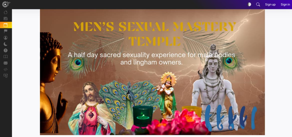
All Posts By Davis Dorothy

Patient-Centered Design: How Advanced Hospital Beds Are Changing Healthcare

By Posted on 4min read6 views

The healthcare industry is always changing. Hospital bed design has become a critical component of this change as we work to improve patient outcomes and experiences. The core of these modifications is patient-centered design, which places an emphasis on total wellbeing, safety, and comfort. This piece will discuss how modern hospital beds are transforming healthcare by placing the patient at the center of the treatment process.

The Evolution of Hospital Beds

Before diving into the advanced features of modern hospital beds, let’s take a moment to appreciate how far we’ve come. Hospital beds have undergone a remarkable transformation throughout history. In the past, they were functional but far from comfortable. Today, patient comfort is a top priority.

Advanced Features of Modern Hospital Beds

Adjustable Positions and Ergonomics

Modern hospital beds mark a notable departure from the rigid, one-size-fits-all structures of the past. These state-of-the-art beds offer a versatile range of adjustable positions, empowering patients to find their optimal comfort. Whether it’s lifting the head for easier breathing or elevating the foot section to boost circulation, these beds are custom-tailored to address the distinct needs of every individual.

Pressure Ulcer Prevention Technology

In the realm of modern hospital beds, one standout innovation is the seamless integration of pressure ulcer prevention technology. These beds are thoughtfully designed with meticulous engineering to efficiently redistribute pressure, effectively addressing the common issue of painful pressure ulcers frequently encountered during extended hospital stays. A pivotal feature of these beds is their proactive approach to promoting skin health, ultimately playing a vital role in enhancing a patient’s overall well-being in a holistic sense.

Integration of Smart Technology

In the 21st century, the age of smart technology has transformed even hospital beds. Numerous cutting-edge beds now incorporate smart features, granting patients the ability to manage their surroundings effortlessly. With a simple touch of a button, patients can fine-tune the lighting, operate the television, and more. These beds empower patients by placing control right at their fingertips.

Enhancing Patient Comfort and Experience

Customization Options for Patients

Personalization plays a pivotal role in patient-centered design. Contemporary hospital beds provide an array of customization choices, including the selection of mattress type and firmness. This high degree of personalization is geared toward ensuring patients experience greater comfort throughout their hospital stay, ultimately leading to a positive influence on their recovery process.

Improved Sleep Quality and Pain Management

Prioritizing the rest patients require is absolutely crucial, and it commences with ensuring they enjoy high-quality sleep. These purpose-built beds surpass the ordinary, elevating the overall quality of sleep and, in turn, promoting the healing process. Packed with a wide array of features, including integrated massage functions and cutting-edge pain management systems, these beds operate harmoniously to craft an exceptionally comfortable and pain-free experience for individuals on the path to recovery and revitalization.

Benefits for Healthcare Providers

Enhanced Patient Care and Monitoring

Advanced hospital beds don’t just benefit patients; they also make life easier for healthcare providers. These beds often come with built-in monitoring systems that allow for real-time tracking of patient vital signs and movements. This technology streamlines care and helps healthcare providers deliver the best possible treatment.

Reducing Workload for Nursing Staff

Nursing staff play a critical role in patient care, but their workload can be overwhelming. Advanced hospital beds can alleviate some of this burden. Features like automated patient repositioning and alarms for potential issues reduce the need for constant manual monitoring.

Real-Life Applications

To truly understand the impact of patient-centered design and advanced hospital beds, let’s explore some real-life applications. We’ll delve into case studies of hospitals that have embraced this innovative approach to patient care.

Challenges and Considerations

While the benefits of advanced hospital beds are clear, there are challenges and considerations to keep in mind. One major concern is the cost. These beds can be expensive, and not all healthcare facilities can afford the initial investment. Additionally, training staff to use the technology and maintaining the beds can be complex.

Future of Patient-Centered Design

The bright prospects for patient-centered design signal forthcoming significant advancements. Expect a multitude of innovations in hospital bed technology, including the seamless integration of electronic health records and telemedicine. It’s clear that patient-centered care is progressively gaining traction, extending beyond the hospital bed. This expansion is poised to drive a profound shift across the entire healthcare landscape, assuring a future where the patient stands at the forefront of healthcare innovation and delivery.

Conclusion

In conclusion, patient-centered design is a game-changer in healthcare. Advanced hospital beds are at the forefront of this movement, prioritizing patient comfort, safety, and overall experience. As we look to the future, it’s clear that the healthcare industry is committed to putting the patient at the center of care, and advanced hospital beds are a significant step in that direction. The days of uncomfortable, one-size-fits-all hospital beds are behind us, and the future looks brighter and more comfortable for patients.

Share

Sports Medicine in Asia: A Technological Revolution for Athlete Wellness

By Posted on 4min read9 views

In the world of sports, where milliseconds can determine victory or defeat, athletes and their teams are always in pursuit of the latest edge. In recent years, a revolution has been brewing in Asia, one that promises to redefine the way athletes prepare, recover, and perform. Welcome to the fascinating world of sports medicine in Asia, where cutting-edge technology, innovative practitioners, and a commitment to athlete wellness have combined to create a transformation like no other.

The Digital Diagnosis

Imagine an athlete straining a muscle on the field. In the past, diagnosing the extent of the injury might have taken hours, if not days. But not anymore. The rise of technology has revolutionized the way we diagnose sports injuries. Advanced imaging techniques such as MRI and CT scans have become as commonplace as Asian Betting Sites, providing instant insight into an athlete’s condition. These digital marvels enable sports medicine professionals to make rapid, precise assessments, leading to faster treatment decisions and quicker recovery.

High-Tech Rehabilitation

Rehabilitation is a critical phase of an athlete’s journey back to peak performance. In Asia, cutting-edge rehabilitation techniques have taken center stage. Robotic-assisted therapies, once seen only in science fiction, are now a reality. Athletes use exoskeletons and robotic devices to rebuild strength and mobility. These innovations offer a level of precision and effectiveness that was previously unattainable, helping athletes return to their sports with minimal downtime.

Wearable Wonder

From smartwatches that monitor heart rate to GPS-enabled shoes that track movements, wearable technology has become an athlete’s best friend. These wearables provide real-time data on performance, helping athletes and their coaches make informed decisions. They not only enhance performance but also play a crucial role in injury prevention. By analyzing an athlete’s movements and vital signs, wearables can detect signs of fatigue or stress, alerting the team to potential issues before they become severe.

Nutrition and Performance

In the quest for peak performance, nutrition plays a pivotal role. Asian sports medicine has witnessed a revolution in this aspect too. Nutritional plans tailored specifically to athletes are now commonplace. These diets are carefully calibrated to optimize energy levels and support muscle recovery. AI-driven dietary planning takes the guesswork out of meal preparation, ensuring that athletes get the right nutrients at the right time.

Game-Changing Surgeries

When injuries require surgical intervention, athletes now have access to minimally invasive procedures that minimize scarring and recovery time. Advances in orthopedic surgeries are allowing athletes to return to their sports faster than ever. These innovations are a testament to the relentless pursuit of excellence in sports medicine.

Asian Pioneers

Behind the scenes of this revolution are visionary Asian sports medicine practitioners. Their groundbreaking research and innovative practices have not only improved the well-being of athletes in Asia but have also influenced the global sports medicine community. These pioneers are at the forefront of discovering new approaches to injury prevention and performance enhancement.

The Impact on Asian Athletics

The impact of this technological revolution on Asian athletics is undeniable. Athletes across various sports have benefited from modern sports medicine practices. From the cricket grounds of India to the martial arts dojos of Japan, athletes have experienced faster recoveries, reduced injuries, and improved overall performance. This has had a positive ripple effect on the region’s sports scene, inspiring more young talents to pursue their athletic dreams.

Challenges and Ethical Considerations

However, with great power comes great responsibility. The increasing use of technology in sports medicine has raised ethical questions. Balancing technological advancements with the human element of healthcare is a challenge. Sports medicine practitioners must navigate the fine line between enhancing performance and maintaining the well-being of the athletes.

Future Horizons

What does the future hold for sports medicine in Asia? Experts predict that artificial intelligence will play an even more significant role. AI algorithms will help predict injury risks, customize training programs, and provide real-time insights into an athlete’s condition. The potential for AI and other technologies to revolutionize athlete wellness is limitless.

 A Global Perspective

The impact of Asian sports medicine is not limited to the continent itself. It is influencing the world. Collaborations between Asian and Western medical professionals have led to a rich exchange of knowledge. The global sports community is increasingly adopting the practices and techniques developed in Asia, further solidifying the region’s role as a leader in the field.

The Role of Government and Industry

Support from governments and the private sector has been instrumental in this revolution. Investment in sports medicine research has led to breakthroughs that benefit both professional and amateur athletes. As governments recognize the importance of athlete wellness, they are allocating resources to ensure that sports medicine continues to advance.

Keeping the Athlete at the Core

Amidst all the technological advancements, one principle remains constant: the athlete is at the core of sports medicine. The athlete-centered approach emphasizes personalized care, tailoring treatments to the individual’s unique needs. This approach ensures that athletes receive the best possible care, enhancing their chances of success and well-being.

Words from the Athletes

To truly understand the impact of this revolution, we turn to the athletes themselves. Their testimonials are a testament to the power of modern sports medicine. Athletes speak of quicker recoveries, reduced pain, and the ability to achieve more than they ever thought possible. Their success stories are a source of inspiration to all.

Conclusion

In conclusion, the technological revolution in sports medicine in Asia has ushered in an era of unprecedented athlete wellness. With state-of-the-art diagnostics, high-tech rehabilitation, wearables, and nutritional advancements, athletes are reaching new heights of performance and well-being. As Asia continues to lead the way in sports medicine, we can only anticipate a brighter future for athletes worldwide. The ongoing revolution in sports medicine shows no signs of slowing down, and the best is yet to come.

Share

Medical 3D printing and its varieties

Medical 3D printing and its varieties

The medical 3D printer has taken on a whole new meaning in recent years due to the rapid development of 3D technology. Now it is a versatile production tool that can be used to perform a wide range of diverse tasks. For example, 3D printing in medicine is actively used for:

Creation of living tissues and organs;
Printing of bones;
Creation of blood vessels;
Manufacturing of biocompatible prostheses;
Creation of individual implants;
Production of unique surgical instruments;
Designing and creating assistive devices and much more.

Of course, these are not the only areas in which 3D printer for medicine is actively used. But these are the areas in which medical organizations worldwide are moving. In many large hospitals and laboratories, 3D printing has already become a very common practice. Professional equipment is purchased en masse for such organizations, but in this section we want to talk more about why you need a desktop 3D printer for medicine.

How a 3D printer for medicine is used
When it comes to the use of 3D technology for medical purposes, many people immediately think of 3D printing of organs and other revolutionary projects. But don’t forget that even ordinary FDM and SLA 3D printers can be of great service to Hippocrates’ followers. Here, in what areas they are useful:

Dentistry. The most common medical industry in which 3D printers are used en masse. Many photopolymer resins and SLA 3D printing devices are even adapted to the needs of dentists. Today, even full-fledged dental instruments and implants can be created using this technique;

Implant manufacturing. Here we are talking about surgical implants, including metal implants. Of course, making a biocompatible titanium implant will require a professional medical 3D printer, but prototypes and master models are made with desktop 3D printing devices;

Custom medical models. Such models are needed for quality medical planning and are most often created from CT scan data. With their help, it is possible to distribute all the stages of treatment quickly and correctly, without disturbing the patient. Roughly speaking, the doctor can perform the operation without surgery;

Orthopedic braces. A very common way to use 3D printers, which requires 3D scanning. Thus, the output is an individual orthopedic corset, which fully corresponds to all anatomical features of the patient.

Creation of training models. A 3D printer for medicine would also be a great companion for medical students and trainees who need to thoroughly study human anatomy. 3D printing is an economical and efficient way to reproduce anatomical models.

Of course, we can not say that every doctor or hospital needs a 3D printer for medicine, but its benefit to this industry is difficult to deny. For example, SLA 3D printing is gradually gaining popularity in the circles of dentists due to its accuracy and convenience.

Share

The misonix bonescalpel ultrasonic bone scalpel

The misonix bonescalpel ultrasonic bone scalpel

Ultrasonic bone dissection with the Misonix BoneScalpel represents a technological breakthrough with unique advantages for spinal surgery. The BoneScalpel allows the surgeon to perform an osteotomy with the efficiency of a sharp chisel, the control of a Kerrison wire cutter, the convenience of a high-speed drill and the thinness of a microsaw, but in the new dimension that ultrasound provides: a highly precise thin cut with reduced bleeding and exceptional tissue selectivity between bone and soft tissue.

The BoneScalpel is unique because it offers a tissue-specific and more precise osteotomy. Its blunted blade makes an effective compression incision in hard bone while remaining atraumatic for soft tissue, which is displaced by the dissipating energy of the blade.

The Misonix BoneScalpel is currently the only ultrasound device that uses blunted blades to frontally plunge into bone, similar to an osteotome. This allows you to perform osteotomies requiring high instrument power with high accuracy and soft tissue safety. The bone can be excised in a single block, including even the dense cortical layer. Ultrasonic blades are widely used for various complex osteotomies to correct various bone deformities in spinal and maxillofacial surgery.

The BoneScalpel shavers allow for layer-by-layer removal of bone, similar to a boron. This allows a gentle removal of bone tissue near delicate structures for decompression, such as in the spinal or radicular canal or near the optic nerve.

Ultrasonic handpieces – shavers perform micromovements in longitudinal direction, which eliminates gyroscopic effect. In contrast to power tools with moving parts, there is no winding or tearing of soft tissues.

Ultrasonic osteotomy significantly reduces bleeding. The BoneScalpel reduces blood loss and therefore the amount of necessary blood transfusions.

ADVANTAGES OF USING ULTRASOUND
The Misonix BoneScalpel is an innovative ultrasound surgical device for fast, safe and precise osteotomies. It is designed specifically to make thin and clean cuts in bone structures with minimal bone loss and preservation of underlying and adjacent soft tissue.

The BoneScalpel receives an electrical signal with a nominal frequency of 22.5 kHz from an ultrasonic generator. Using a piezoelectric transducer, this signal is converted into mechanical vibrations, which in turn are amplified to effective parameters. The blunted BoneScalpel blade makes reciprocating micro-movements in the longitudinal direction, which enables effortless dissection of the hard cortical bone layer.

Ultrasound osteosurgery is tissue-specific, because it makes it possible to precisely cut or remove dense bone tissue without damaging the elastic soft tissue. The loss of bone viability is minimized, and bleeding during surgery is significantly reduced, so that the operating field will remain free of blood. All of this leads to a significant reduction in surgical time.

The BoneScalpel makes the bone cut without damaging the soft tissue, because the bone is more dense, and because of the unique design of the ultrasound instruments.

When the BoneScalpel blade comes into contact with the bone, the bone is not bent, deformed or displaced.

As a result, the bone absorbs a large amount of energy from the blade and is destroyed at the point of contact.

By contrast, soft tissue, due to its elasticity, comes into contact with the BoneScalpel blade and shifts, deforms and vibrates.
As a result, a significant portion of the energy is dissipated and damped in the tissue.

Usually the amount of energy absorbed by the soft tissue at the point of contact with the blade is not enough to cause any damage to the soft tissue. The exception to this is when contact with the blade causes tissue tension and this contact occurs over a long period of time.

INDICATIONS FOR USE AND CLINICAL EXPERIENCE
The BoneScalpel system is indicated for cutting and fragmenting bone tissue as well as for treating wounds (such as burns, trophic and diabetic ulcers, bedsores, etc.) and soft tissue in the operating field where the doctor believes a powerful ultrasound disintegrator is required.

Share

What are the laboratory devices

What are the laboratory devices

Vacuum systems (tubes)
Vacuum systems (tubes) are used for blood sampling for analysis. Modern production technology guarantees high quality of the finished product. The device is disposable and can be used to collect the required amount of blood quickly and safely.

Thanks to the vacuum, the specialist can take a certain amount of material, observing the dose. The blood can be stored in the tube for some time and transported if necessary. All these aspects allow for error-free testing, correct diagnoses, and help people recover from serious illnesses.

Each test tube has a color marking that has its own special meaning. It indicates the purpose of the equipment for a particular type of analysis. The tubes are hermetically sealed, do not allow air to pass through, and do not react to shocks and mechanical damage. With their help, it is possible to store the material in a frozen state for some time.

Portable venous scanner
Medical specialists know that if a needle is inserted incorrectly, a patient may experience complications, deterioration of health, and the development of tissue necrosis. In particularly serious and complicated cases, you can’t take any chances. If you are looking for high-quality laboratory instruments and equipment, pay attention to the portable venous scanner that you can purchase from us. It allows you to perform venipuncture in dangerous places – near the eyes, on the temple and neck, and to administer intravenous injections.

This device is usually used to treat patients with cancer, drug addiction, increased body hair growth, overweight, etc. The equipment allows detecting and diagnosing varicose veins and subcutaneous bleeding.

Laboratory centrifuges
Centrifuges are standard equipment in every laboratory and are intensively used to separate components of different densities. The company offers a wide range of laboratory centrifuges, rotary and tilt rotors, fixed-angle rotors, cytology and hematocrit rotors, rotors for microtiter plates, as well as a wide range of adapters of various configurations. Thanks to the possibility of supplementing rotors and beakers with adapters, the user can work with containers of different structure and volume, which makes the centrifuge operation process convenient and simplifies it as much as possible. All products meet the highest standards of reliability and safety.

Share

What are modern CT scanners like?

By Posted on 3min read0 views

What are modern CT scanners like?

Thanks to the modern CT scanner, medical professionals can now examine every part of the human body. After the procedure, the computer device displays the result on the monitor screen in the form of a 3D model of the scanned organ, plus a complete analysis of it.

The main advantage of this type of equipment is: high quality of images and their processing. Therefore, tomographs are indispensable in all branches of medicine.

There are three types of such devices in the medical market:

– magnetic resonance imaging;
– multislice computed tomography;
– Positron or two-photon emission.

Magnetic resonance imaging (MRI), in turn, come in three types: closed, open and mobile. MRI machines differ in the type of magnets used, namely:

– equipment with permanent magnets made of ferromagnetic alloys;
– device with a resistive electromagnet (this is a solenoid made of iron and copper wire);
– models with superconducting electromagnets (solenoid of niobium-titanium alloy).

Computed tomographs are also found in three types:

– single-slice spiral (the surface with the sensors and the radiation source move synchronously in the process);
– with two X-ray tubes (makes the clearest tomogram of the heart, each tube works in autonomous mode);
– multilayer (has several rows of receiving sensors, makes it possible to examine the entire organ).

Each type of CT scan may differ from each other according to the following parameters: speed of full rotation, type of moving part of the device, absence or presence of oblique slices. The most advanced CTs can scan at three revolutions per second. Certain models may have options to provide comfort and a quiet environment for patients. Children are provided with cartoons to watch during the examination, and people with poor hearing are usually repeated voice commands.

Open CT scanners have more open space and a smaller work area surface, which helps patients go through the procedure more relaxed. But they are equipped with a lower magnetic field, the examination time increases and the quality of the images wants to be better.

Closed models of tomographs are characterized by a closed working space, in general the patient is entirely in the equipment, there are openings only at the feet and head of the patient, which sometimes has a negative effect on people with an anxious psyche. They have a high power of magnetic field, it reduces the examination time and produces images of high quality.

Mobile models of this equipment allow the examination to be carried out at any place. The design is a container inside which the tomograph is located, with the container on a mobile platform that is transported to any location.

The MRI image is obtained thanks to radio impulse transmitters, which generate waves of resonant frequency and convert them into pulses of the required shape, and the receiving coil is a sensitive antenna, which is located perpendicular to the direction of the main magnetic field.

Share

Modern diagnostic equipment

By Posted on 1min read0 views

Modern diagnostic equipment

One of the current trends in the development of diagnostic equipment is the development of devices and instruments that can be used by patients who have no medical training. Various portable devices are used at home when a person needs to pay constant attention to his or her body.

Portable glucose meters.
People diagnosed with diabetes mellitus must periodically measure blood sugar levels in order to feel well. Therefore, to purchase a glucometer is the natural desire of each of them. Constantly going to the clinic for tests, especially for the elderly, is very difficult.

A glucose meter is exactly the device, which allows to take measurements in any conditions.

Electrocardiographs
Electrocardiographs also belong to the class of diagnostic medical equipment. They record electrical potentials that occur in the heart and display them as an electrocardiogram.Electrocardiograph

This device records such indicators

such as the regularity of the heartbeat,
The rate of heartbeat and some other parameters.

It is used to diagnose cardiovascular diseases.

Portable cardiographs can be used under any conditions and do not require any preliminary preparation of the patient.

Share

Principle of operation of ultrasonic devices

By Posted on 2min read0 views

Principle of operation of ultrasonic devices

Today, ultrasound scans are one of the most popular medical diagnostic methods. Ultrasound scanners are firmly established in every modern clinic, diagnostic room and even intensive care ambulance. The basic principle by which to classify ultrasound machines, is the scope of their application, and, depending on this, they are stationary and portable.

But whatever ultrasound machines, they all work on the same principle and, in general, have a similar internal structure.

The construction of ultrasound scanners
The main elements and units of the ultrasound machine are:

ultrasound transducer – the most important part of the scanner, designed for the formation and reception of sound waves;
pulse control sensor – involved in changing the strength, frequency and duration of pulses transmitted by the ultrasound transducer;
Processor unit – a computer designed to receive and process pulses from the transducers with subsequent output of human-understandable information to the monitor;
monitor – visualizes the data received from the processor unit;
keyboard, trackball (mouse) – allows entering initial data, as well as changing or adding data during or after the diagnostic session;
printer – designed for saving the obtained data on paper.

Ultrasound scanner operation is based on the properties of sound waves generated in the ultrasound sensor using the piezoelectric effect. Sensors differ in shape and size, which determine the area of visibility, as well as the operating frequency, which determines the depth of sound wave penetration (depth of scanning) and the resolution of the data obtained (picture quality). Sensor type will depend on the type of transducer, scanning method, and currently there are 4 types of ultrasound sensors: sector mechanical, linear, convex and mini-convex, phased sector.

The heart of the sensor is several quartz piezoelectric crystals which, if exposed to an electric current, begin to vibrate and emit a sound wave. When a reflected sound wave hits the crystals, they are able to produce an EMF. These unique properties of quartz crystals have led to the creation of ultrasound scanners and a whole new field of medical diagnostics. To filter the sound waves hitting the sensor, it is equipped with a special filtering layer, and an acoustic lens allows you to tune in to receive the desired wave.

How it works
The principle of operation of any ultrasound machine is as follows. The sensor emits sound waves of varying frequency (from 1 to 18 MHz), amplitude and duration. Part of the waves, penetrating into the body and reaching the boundaries of media (e.g., liquid – soft tissue or soft tissue – bone), penetrate further, and part – is reflected and recorded by the sensor. Based on the known speed of sound propagation in certain media of the body, the processor unit calculates the time of sound wave passage from the sensor to the scanned organ, which as a result allows you to make a general picture of the diagnosis and draw conclusions.

Share

Lasers for excimer laser vision correction

Lasers for excimer laser vision correction

Amaris 500E Excimer Laser (Schwind, Germany)
Sshwind Amaris 500E Excimer Laser is a state-of-the-art technological innovation in the field of laser vision correction systems. Numerous clinical trials have proved high efficiency and safety of Amaris 500E for improvement of visual acuity and quality of vision in myopia, hyperopia, astigmatism and higher degree aberrations (refraction distortions).

Automatic Fluence Level Adjustment “AFLA” (Automatic Fluence LevelAdjustment) provides optimal correlation between the number of laser pulses and the laser energy delivered to the tissue, resulting in quick ablation with optimal corneal topography alignment. The laser system with a 500 Hz laser pulse repetition rate is efficiently supplemented with a 5D eyetracker which compensates for micro movements of the eye in five dimensions. You will get the best quality of correction, the procedure will be safe, and rehabilitation will be fast.

IntraLase FS60 femtosecond laser (USA)
This femtosecond laser is used to perform excimer laser correction according to a revolutionary femtolaser technique. In femtosecond laser eye correction corneal flap is formed not with a microkeratome but with the help of femtosecond laser beam, i.e. it is absolutely noncontact. IntraLase FS60 femtosecond laser has high frequency and short pulse duration.

Pulse duration is measured in femtoseconds (one trillionth of a second, 10-15 s), which allows to separate corneal layers at molecular level without heat generation and mechanical effect on surrounding eye tissues. The femtosecond laser beams are focused at a preset depth with an accuracy of a few microns to create a layer of “micro-bubbles” of the desired configuration. By positioning multiple micro-bubbles in the neighborhood using a computer-controlled program, the ophthalmic surgeon is able to obtain a plane of separation of absolutely any shape with the highest precision.

The corneal flap formed in the process of femtosupported laser correction is uniform in thickness, thin and has smooth borders. The constant thickness of the corneal flap excludes higher order aberrations due to the effect of inhomogeneous refraction of rays, increasing safe procedure and dramatically reducing rehabilitation period. The ideal smooth surface of the corneal tissue separation border enables to achieve a significant reduction in the rehabilitation period and preserve high contrast sensitivity.

IntraLase FS60 femtosecond laser is a member of the complete line of iLasik system. It works in combination with Amaris 500E excimer laser and WaveScan aberrometer. This complex makes it possible to perform laser vision correction taking into account the smallest peculiarities of a patient’s visual system.

Share

Vacuum laboratory equipment and centrifuges

Vacuum laboratory equipment and centrifuges

Vacuum equipment is often used in laboratories, especially those performing electrophysical research.

Vacuum equipment includes:

pumps;
installation systems;
chambers.

It is possible to choose the necessary type of vacuum equipment based on the tasks set for the laboratory.

You may need to buy centrifuges and sieves. Laboratory centrifuges are devices that are used to separate solids from liquids or substances with different specific gravities.

Most commonly used in medical and industrial laboratories, they are also used in chemical and biological experiments.

Centrifuges have a lid with a twist-off screw, which is used to close the drum tightly. When the device starts to work inside it, a centrifugal force is generated due to the high speed of rotation, which separates the substance into particles. They are sifted through special sieves, light substances move to the center, and heavy particles settle at the bottom.

When working, employees will need sterilizers and sterilization indicators. This is especially true in medical laboratories. Vacuum steam sterilizers or gravity sterilizers are most commonly used. Chemical sterilization indicators are also used to monitor sterility.

When dealing with liquids and aqueous solutions in laboratories, distillers and bidistillers are used.

Share